RAD 23N: Seeing the Invisible
General Education Requirements
Course Description
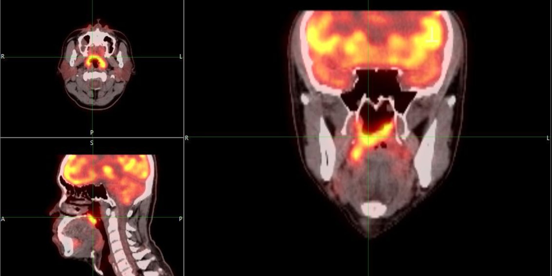
Seeing is believing! Of all the possible photon wavelengths out there, however, only a small range of about 380 to 720 nanometers – known as the visible spectrum – can be detected by our light-sensitive cone cells. Scientists have been developing technologies that help overcome this limit. Optical microscope was first used in the 17th century to see microorganisms; Magnetic resonance imaging (MRI) allows non-invasive visualizing of human internal organs. This seminar course will introduce these breakthrough imaging technologies from super resolution fluorescence microscopes for imaging single molecules in living cells to MRI and positron emission tomography (PET) for visualizing the happenings deep inside our bodies. We will learn about their applications in probing physiology, biology and biochemistry for biological research and medical diagnosis. Students will have the opportunity to tour an imaging facility and perform hands-on laboratory imaging. Finally, students will identify an imaging problem, research possible solutions, and present it to the class.
Meet the Instructor: Jianghong Rao

“I am a professor in the Department of Radiology with a courtesy appointment in the Department of Chemistry. I developed my passion about molecular imaging during my postdoc at UCSD where I learned live cell imaging. I was excited about the potential of molecular imaging to revolutionize the way we diagnose and treat diseases, and in 2004, I joined the newly formed Molecular Imaging Program at Stanford to continue my interest in this field. My lab works on designing and building molecular tools to image and interrogate diseased cells in our bodies, such as new methods for detection of bacterial pathogens, new ways to label and track cancer cells, and new nanomaterials to selectively kill cancer cells.”


