General Education Requirements
Course Description
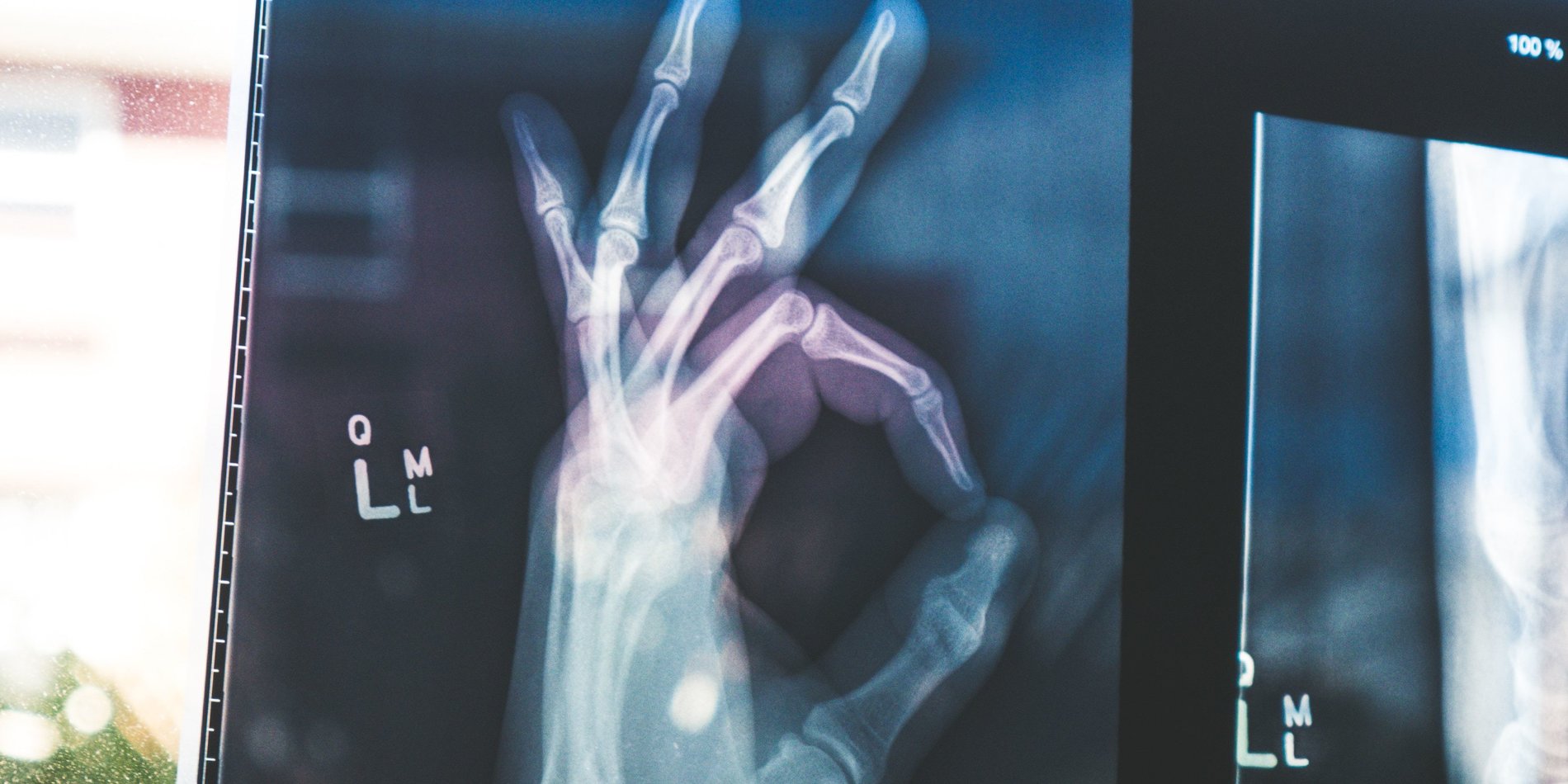
For centuries, the only way to know what was happening inside our bodies was to open them up, and look. All that changed very late in the 19th century and throughout the 20th century, with the development of increasingly potent tools such as X-ray, computed tomography (CT), magnetic resonance imaging (MRI), positron emission tomography (PET) and ultrasound.
Today, X-rays can depict tiny bone cracks, ultrasound can visualize heart valve dysfunction, CT can map our vascular system, MRI can see small brain defects, and PET imaging can help identify aggressive cancers.
In this seminar, we will discuss the magic of medical imaging and the principles and technologies behind these tools that enable seeing inside our body. We will cover the main medical imaging modalities, and discuss their applications with real-life examples.
Students will learn about medical imaging as well as about common conditions and diseases, and aspects of human anatomy. Essential components of the seminar include active participation during the discussions and student-led presentations on medical imaging topics of interest.
The seminar has no prerequisite other than an interest in medical imaging and curiosity about the human body.
Meet the Instructors: Daniel Ennis & Marios Georgiadis
Daniel Ennis

“I am a Professor in the Department of Radiology in the School of Medicine. My early scientific interests were at the intersection of engineering and medicine. This led me to pursue my undergraduate studies in bioengineering at UCSD, followed by my Ph.D. graduate work at Johns Hopkins in biomedical engineering. My interest in medical imaging arose from a fundamental interest in cardiac performance. I now oversee the Cardiac Magnetic Resonance Group wherein we invent, develop, validate, and deploy new MRI techniques that help evaluate cardiac structure, function, flow, and remodeling in pediatric and adult patients. We also have an interest in mapping the billions of cardiomyocytes that make up the heart and enable us to be active and athletic humans.”
Marios Georgiadis

“I am an Instructor in the Department of Radiology in the School of Medicine. With a mechanical engineering background, I have always been intrigued by how our body works, but also by what tools we can use to understand what is going on inside it. After highschool, I was undecided about whether I wanted to be an engineer or a medical doctor; it turns out that these two can almost be combined, in biomedical engineering. As a biomedical researcher, I have used most medical imaging modalities to study bones, muscles, tendons, and the brain, from subcellular to whole organ scales. I am currently studying how our brain changes with old age, neurodegeneration, or after it sustains an impact (e.g. during high-contact sports). At the same time, I am very interested in mapping the billions of connections within our brain, which give rise to our thoughts, our feelings, and our personality.”


